Phone: 0884-2944466,7416622540
vascularsurgeonkakinada@gmail.con
1st floor, D.No: 21-1-10, Jawahar Street, Opp Shravani ENT Hospital, Kakinada-533001.
Phone: 0884-2944466,7416622540
vascularsurgeonkakinada@gmail.con
1st floor, D.No: 21-1-10, Jawahar Street, Opp Shravani ENT Hospital, Kakinada-533001.
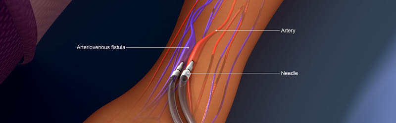
An arteriovenous fistula is a medical condition where an artery and vein connect directly, causing blood to flow between them. It can happen at virtually any place in your body where an artery and vein are close together, especially inside of your organs and limbs (arms and legs). Depending on where fistulas are, why they happened and their size, they can be harmless (or even helpful when there’s a medical reason to create one), or they can be a major health issue and permanently damage your heart.
Arteriovenous fistulas (pronounced “are-tee-re-oh-vee-nus fis-tew-las”) can happen to anyone at any age, especially when they happen because of injuries. Certain types of fistulas are more common in certain groups of people, usually because of their life circumstances
To diagnose an arteriovenous fistula, a health care provider may use a stethoscope to listen to the blood flow in the arms and legs. The blood flow through an arteriovenous fistula makes a sound like humming
Duplex ultrasound. Duplex ultrasound is the most effective and common way to check for an arteriovenous fistula in the legs or arms. In duplex ultrasound, sound waves are used to evaluate the speed of blood flow.
Computerized tomography (CT) angiogramThis imaging test can show if blood flow is bypassing the capillaries. Dye (contrast) is given by IV for this test. The dye helps blood vessels show up more clearly on the images.
Magnetic resonance angiography (MRA)This test may be done if you have signs of an arteriovenous fistula deep under the skin. Like an MRI, an MRA uses a magnetic field and radio waves to create pictures of the body's soft tissues. Dye (contrast) is given by IV to help blood vessels show up better on the images.
If an arteriovenous fistula is small and doesn't cause any other health problems, close monitoring by a health care provider may be the only treatment needed. Some small arteriovenous fistulas close by themselves without treatment.
Ultrasound-guided compression. This may be an option for an arteriovenous fistula in the legs that's easily seen on ultrasound. In this treatment, an ultrasound probe is push down on the fistula for about 10 minutes. The compression destroys blood flow to the damaged blood vessels.
Catheter embolization. In this procedure, a thin, flexible tube (catheter) is inserted in an artery near the arteriovenous fistula. Then, a small coil or stent is placed at the site of the fistula to reroute blood flow. Many people who have catheter embolization stay in the hospital for less than a day and can resume daily activities within a week.
SurgeryLarge arteriovenous fistulas that can't be treated with catheter embolization may require surgery. The type of surgery needed depends on the size and location of the arteriovenous fistula.